News
Postdoc George Hoeferlin joined the lab
March 1, 2024
New collaboration with Dr. Chen Yang from BU
January 16, 2024
Associate Professor of Neurosurgery
Director, Neural Prosthetic Research Laboratory
Department of Neurosurgery
Massachusetts General Hospital
Harvard Medical School
Shelley Fried, PhD, is the Director of the Neural Prosthetic Research Laboratory and an Associate Professor in the Department of Neurosurgery at the Massachusetts General Hospital. His research focuses on the interactions between artificial stimulation (delivered mostly by implantable prostheses) and neurons of the CNS. Most of his work focuses on visual prostheses within the retina, thalamus, and primary visual cortex. His work strives to better understand fundamental principles of sensitivity to artificial stimulation with a focus on developing strategies to more effectively stimulate non-working portions of the CNS, i.e., better re-create the patterns of neural signaling that arise naturally in the healthy system. Current projects include visual prostheses efforts in the retina, lateral geniculate nucleus, and primary visual cortex, using stimulation modalities that include conventional microelectrodes, high-frequency stimulation, magnetic stimulation from microcoils, and optoacoustic stimulation. In addition to neural prostheses efforts, the lab also studies the spike initiation mechanism in retinal ganglion cells.

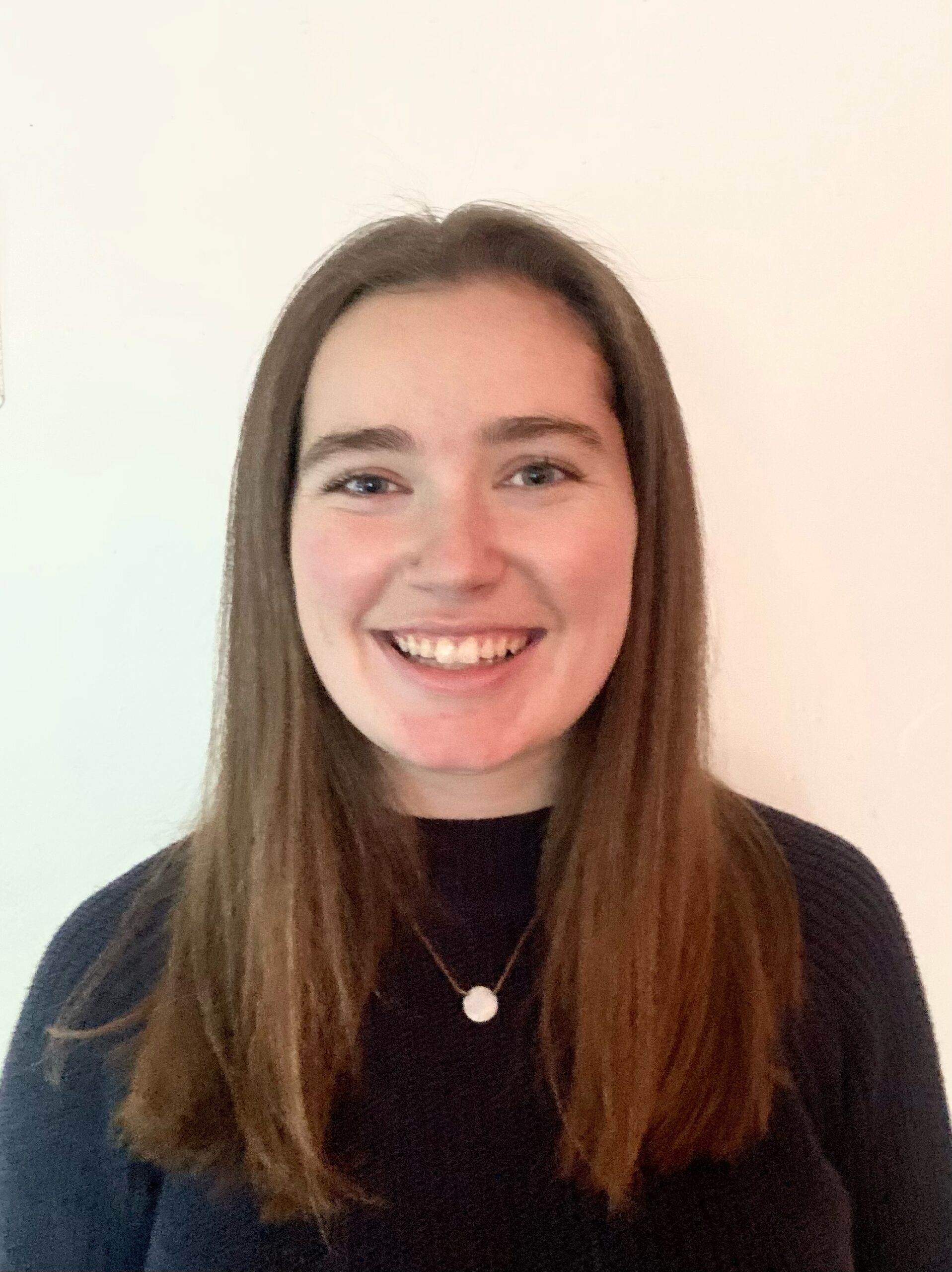
I am Kaitlyn Kessel and I am originally from Rockport Maine and moved to Boston 4 years ago. I graduated from Simmons University in May 2023 with a Bachelor of Science in Biology. During my undergraduate journey, I had the privilege of being recognized as a SURPASs Grant recipient and a proud member of Sigma Xi. My passion for research led me to explore the intriguing world of biology where I ended up doing a thesis specialized in environmental toxicology. Throughout my undergraduate career, I was also a teaching assistant where I was dedicated to helping students dive into the exploration of the scientific world with hands-on experience
Structural changes in axon initial segments of ON-sustained alpha retinal ganglion cells in retinal degenerated mice. I am actively engaged in a collaborative project alongside Molis, focusing on the comparative analysis of retinal components in both wild-type and RD (photoreceptor-degenerated) mice. Our primary objective is to investigate alpha retina ganglion cells in these two mouse groups. Specifically, we are imaging the Axon Initial Segment (AIS) and Nav1.6 channels on the confocal microscope. Our research delves into the eccentricity of each ganglion cell in relation to the lengths of the AIS, Nav1.6 channels, and axon hillock. These factors play pivotal roles in shaping the dendritic field diameter and soma diameter of each imaged cell. Through this project, we aim to unravel the intricate relationship between retinal neuronal components and their functional significance, particularly in the context of photoreceptor presence or absence.

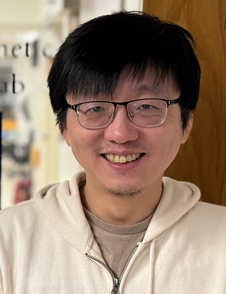
I am an instructor working in the laboratory of Dr. Fried. My primary research focus centers on the development of innovative stimulation strategies for implantable neural prosthetic devices.
Investigating the response of CNS neurons to high-frequency electric stimulation. Retinal prostheses are a promising solution for individuals suffering from outer retinal degenerative diseases, such as macular degeneration and retinitis pigmentosa. However, the quality of restored vision remains limited, likely be due to the mismatch between artificial and natural neural signaling. A stimulation strategy employing high frequency stimulation (~2 kHz) offers potential to selectively target individual retinal cell types, enabling better replication of natural signaling. My project focuses on understanding the mechanism underlying this selective activation through physiological and anatomical studies, aiming to enhance artificial vision quality.
Development of a micro-coil based cochlear implants
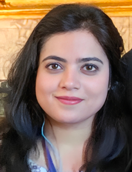
: Cochlear implants (CIs) enable speech recognition for those with severe hearing loss, but performance is limited in noisy environments and most users cannot appreciate music. Suboptimal performance arises because existing devices typically create only ~10 independent spectral channels, much less than that utilized by the healthy cochlea. I have tested a feasibility of a magnetic stimulation from microcoils for cochlear implants. The preliminary results showed that the magnetic stimulation creates narrow channels, i.e., much narrower than those created by the electrodes used in existing CIs. This offers a way to increase the number of independent channels and therefore opens new and exciting possibilities for improving the quality of artificial hearing.
Lee, J. I., Seist, R., McInturff, S., Lee, D. J., Brown, M. C., Stankovic, K. M., & Fried, S. (2022). Magnetic stimulation allows focal activation of the mouse cochlea. Elife, 11, e76682.

AIS projects: The axon initial segment (AIS) is a portion of the proximal axon responsible for spike initiation and back propagation. It is most sensitive region of the entire neuron to artificial stimulation. The AIS structural properties, such as length and distance from the soma, and the distribution of various voltage-gated channels, such as sodium and potassium channels, are tailored to optimise the input/output properties of the neuron and can be altered in response to changes in network activity. Therefore, understanding the AIS properties in healthy and diseased visual system is essential in improving the sensitivity to artificial stimulation. Using immunolabeling followed by confocal microscopy, we can visualise the AIS structure and the voltage gated channel distribution. Paired with in vitro whole-cell patch recording, we are able to relate the AIS properties to the spiking responses of individual neurons.
LGN project: The lateral geniculate nucleus (LGN) is a multilayered structure in the thalamus that receives projection from the retinal ganglion cells before relay the visual information to the primary visual cortex. Compare to retina and the cortex, which are the two most popular targets for visual protheses, LGH offers several important advantages. It bypasses the retina entirely for patients whose retinas are not viable for prosthesis. On the other hand, the neural coding within the LGN is fairly similar to that of the retina and much less abstract than that of the cortex. In this project, we are evaluating the effects of stimulation of the LGN, focusing on how specific stimulation strategies influence the resulting responses in the visual cortex. In parallel, we are characterizing the AIS properties of different types of LGH neurons to understand how such properties influence the cell’s response to stimulation.

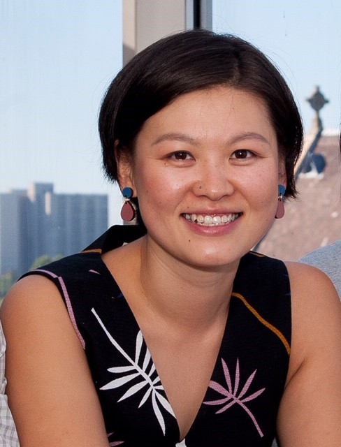
Hannah Rana is a Schmidt Science Fellow in the Fried Lab working on models that artificially simulate the neural code of the healthily operating and the degenerated retina, using recorded electrical spiking from retinal samples measured using Multi-Electrode Arrays (MEAs) whilst subjected to varying light and electrical stimuli, in collaboration with Prof. Zeck’s biomedical devices group at TU Wien. The ultimate aim of these efforts is to reconstruct an artificial retina, which can be used to inspire the design of a physical retinal prosthetic device for vision restoration for the blind. In a second project, she works on spike sorting and signal processing of data output from MEA chips implanted onto the visual cortex of blind subjects, performed by Prof. Eduardo Jover’s lab at the Universidad Miguel Hernandez Biomedical Engineering center. The aim of this work is to correlate the neural activity in the visual cortex with visual perception. She pivots her knowledge gained in developing instrumentation for astrophysics to solving neural detection problems in the human retina and visual cortex. Her interests include applying machine learning, deep learning, computer vision and optimization methods to neural coding and circuitry, detector devices, and instrument design for vision restoration applications.