We are developing a prosthesis that targets the lateral geniculate nucleus (LGN) of the thalamus as a means to restore vision to the blind. The LGN is an attractive site for an implantable prosthesis because it is spatially more expansive than the retina and thus enables higher acuity and a larger number of stimulation sites. At the same time, the neural signaling patterns used by LGN neurons are much less abstract than those of the visual cortex, thereby allowing for more straightforward encoding schemes with the prosthesis. While it has been challenging to develop a high-count, multi-channel device that can safely be implanted into a deep brain structure, the use of splaying electrodes overcomes these concerns. A functional LGN prosthesis is particularly attractive as it would be suitable for just about all blind patients.
Background
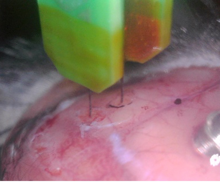
Vision restoration efforts that target the retina are not useful for some of the more prevalent blinding disorders such as diabetic retinopathy and glaucoma, or any other disorder in which viability of the neural retina and/or the pathway to the brain (optic nerve) are lost. In most of these cases, the LGN is intact and thus offers a target for stimulation with the potential for treatment for almost all causes of blindness. Although targeting the visual cortex similarly provides a treatment option to larger numbers of blind subjects, there are some distinct advantages associated with targeting the LGN. For example, the neural coding used within the LGN is simpler than the abstract and spatially distributed coding used in cortex, and thus easier to re-create with a prosthesis. Further, the relatively large, laminated and visuotopically-organized 3-dimensional structure of the LGN facilitates the selective targeting of physiological sub-classes of neurons further facilitating the re-creation of physiological signaling patterns. Another favorable feature of the primate LGN is the interleaving of input from the two eyes, with LGN layers 2, 3 and 5 receiving ipsilateral input and layers 1, 4, 6 receiving contralateral input. From a visual rehabilitative standpoint, implantation into one LGN would provide vision to the contralateral visual field of both eyes, which is not possible with retinal implants. Bilateral implants could provide a wide field of vision to both eyes, which would be valuable for object avoidance when navigating. Further, the central-most LGN cells have receptive fields of 2 minutes of arc (i.e., 1/30 of 1° of visual angle), just above the resolution required for “normal” (i.e., 20/20) vision. The LGN therefore provides a neuronal substrate that allows for both wide-field and high-resolution vision. Splaying multi-electrode arrays (MEAs) enable broad coverage of regions deep within the brain. The device is inserted into a cannula as it advances through the cortex and other peripheral regions of the brain, i.e., to prevent splaying as the device passes through outer brain regions. Once close to the LGN, the MEA is advanced, gradually emerging from the cannula and begins to splay as it comes in contact with parenchyma. Controlled spreading of individual tines of the MEA results in good 3-D coverage of the LGN.
What We Do
There are many gaps in our understanding of how to effectively stimulate the LGN with a prosthesis. We are advancing understanding by studying how LGN neurons respond to electric stimulation with the goal of developing stimulation strategies that effectively drive the LGN. Efforts include in vitro and in vivo experiments in both wild-type and retinal degenerate mouse models. We are learning how to optimize activation for different cell types within the LGN and then assessing how such strategies influence propagation to primary visual cortex. As part of this effort, we are collaborating with multiple groups developing splaying LGN implants and helping each to optimize their design.